Small-angle scattering techniques offer new insight towards the treatment of Alzheimer’s disease

Aggregates of amyloid beta- (Aβ-)peptide, known as fibrils, are one of the hallmarks of Alzheimer’s disease and play a key role in the sequence of events leading to dementia symptoms. Using small-angle neutron and X-ray scattering, researchers from Lund University and the Paul Scherrer Institut have determined the detailed structure of Aβ42-fibril, obtaining important information to design future therapeutics.

Advances in medicine and improved global living standards mean that people are living longer than ever before. However, with the global population ageing at an unprecedented rate, geriatric diseases, including neurodegenerative disorders, have become increasingly prevalent. Alzheimer’s disease (AD) and other forms of dementia are now among the top 10 causes of death worldwide and with no effective treatments currently available, their prevention and cure are a global health priority.

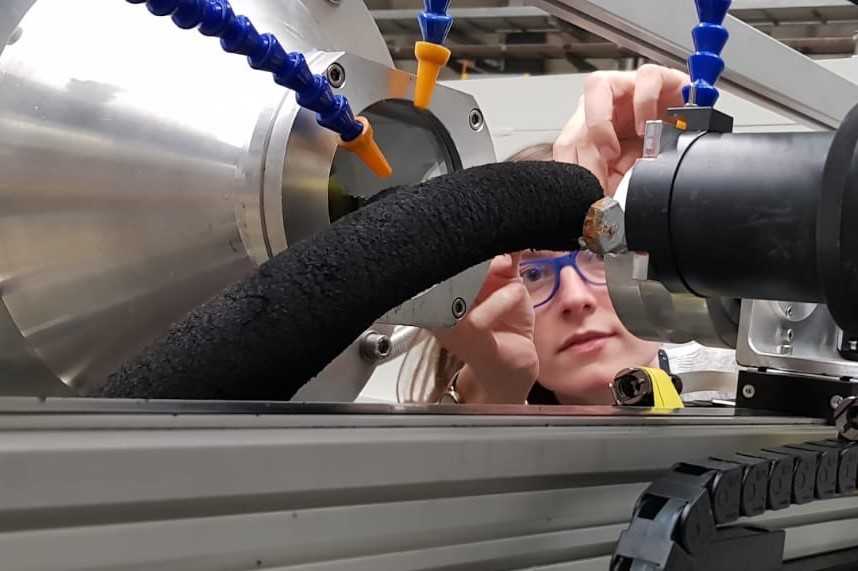
Veronica Lattanzi, a researcher from the Department of Biochemistry and Structural Biology at Lund University, loads a sample of Aβ42-fibrils on the SANS-II instrument at the Swiss Spallation Neutron Source (SINQ).

Growing evidence has linked the onset and progression of AD to the aggregation of Aβ-peptide, of which the Aβ42 variant is most commonly found in AD patients’ brains. Aβ42-peptides can self-assemble into insoluble fibres, known as amyloid fibrils, which catalyse the formation of toxic molecules that damage the brain. Consequently, Aβ42-fibrils are an important target for therapeutics against Alzheimer’s disease.

A team of scientists from Lund University have been working to decipher the atomic structure of Aβ42-fibrils, which will help in designing molecules that can bind to the fibres and inhibit their catalytic activity. In an article published in PNAS, the researchers undertook small-angle neutron scattering (SANS) at the Swiss Spallation Neutron Source (SINQ) and small-angle X-ray scattering (SAXS) to obtain information about the nanostructure of the fibrils. Data from both SAXS and SANS were consistent with a two-filament model, consisting of two twisted stacks of folded Aβ42 molecules. The authors propose that this structure represents a thermodynamically stable state, arising through mainly hydrophobic interactions, as well as from the twisting of the two filaments due to chirality.

Lead author, Veronica Lattanzi, commented that “X-ray and neutrons scattering methods are complementary and versatile methods that can provide new insights to better understand incurable pathologies such as Alzheimer’s disease.”

As well as demonstrating the efficacy of small-angle scattering in the study of biomolecular nanostructures, the results provide key insights to design drugs that control the production of neurotoxic molecules and thereby slow or halt cognitive decline in AD patients.

Trả lời

Email của bạn sẽ không được hiển thị công khai. Các trường bắt buộc được đánh dấu *

Gọi điện cho tôi Gửi tin nhắn Facebook Messenger
Gọi ngay Form Liên hệ Messenger