Small-Angle X-ray Scattering (SAXS) is an invaluable tool for the analysis of liposomes. Liposomes are a class of nanoparticles which are characterized by their phospholipid bilayer wall. Their versatility, ease of manufacturing and biocompatibility makes them attractive carriers for active ingredients in pharmaceutical, cosmetic and food applications. As a consequence, liposomes are among the few classes of nano-based drug delivery systems. SAXS is an efficient tool to probe the size and shape as well as the membrane flexibility and incorporation of active components into the membrane. Moreover, SAXS gives statistically relevant results and can give some unique insights into the liposomes that are not accessible by other techniques.
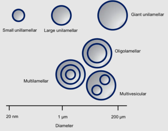
Liposomes, first described in the early 60s, are spherical vesicles with lipid bilayer walls mainly consisting of phospholipids. Their size varies from tens of nanometers to over 200 micrometers. There are two types of liposomes: unilamellar and multilamellar. Multilamellar vesicles are further categorized into multilamellar, oligolamellar and multivesicular liposomes (see the following figure). The relative ease of manufacturing and their biocompatibility renders them an ideal carrier for active pharmaceutical ingredients (APIs) as well as in cosmetic [1] and food formulations. [2]
Apart from various types of phospholipids, liposomes often contain cholesterol or other lipophilic molecules. Furthermore, the surface can be modified with polyethylene glycol (PEG) or other polymers, proteins, small molecules, carbohydrates and other moieties to modify their stability, solubility or direct them to the tissues where they should unload their active cargo. This versatility has led to their success as nano drug-delivery systems, which is illustrated by the liposomes that are currently on the market in clinical formulations.[i]
Some examples of successfully marketed formulations are Doxil®, LMX-4® and DepoDur®. What these registered medicines have in common is that the active ingredient is too lipophilic to be administered directly. Therefore, they are encapsulated in a liposomal carrier.
Characterizing lamellarity and lipid bilayer thickness with SAXS
To safeguard the health of consumers and to maintain a constant quality, the production of liposomes for these applications is regulated under Good Manufacturing Practice (GMP) guidelines. These guidelines state that manufacturing processes must be clearly defined and controlled, and that any changes in the process must be evaluated.[ii] Morphological analysis is a crucial part of the quality control and Small-Angle X-ray Scattering is a well-suited tool for this.
Lamellarity, i.e. the amount of lamellar bilayers, is one of the parameters the FDA (Food and Drug Administration) marks as important. The lamellarity is a predominant factor in the ability of the liposome to contain and retain the drug substance.[iii] The number of lipid bilayers and their exact conformation can easily be accessed with SAXS. Coupling the measurement with Wide Angle X-ray Scattering (WAXS), information about Alkyl chain spacing can be obtained. This allows the determination of gel, lipid or ripple phases.
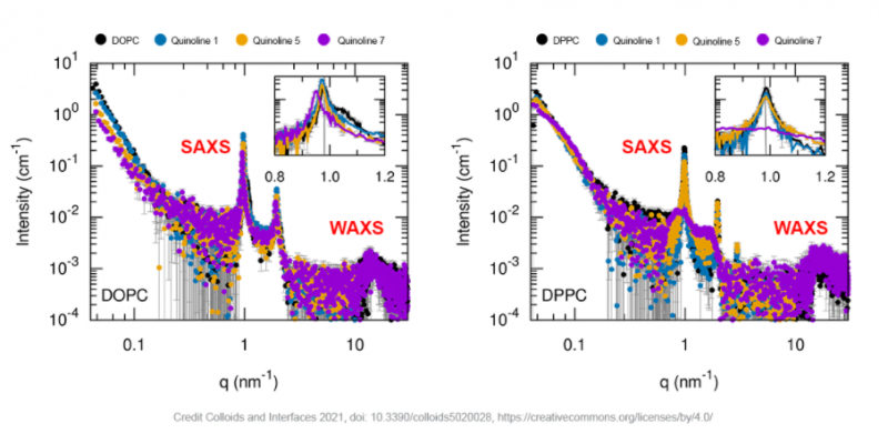
As an example, in a recent publication from scientists from Universita degli Studi dell Aquila and University of La Sapienza in Italy, SAXS was used together with other characterization methods to study the loading of Quinoline in liposomes composed of different lipids [3]. Quinoline is a well-known, and versatile compound, however with low solubility limiting its impact for pharmacological applications which can be overcome with its inclusion into liposomes. The figure below shows SAXS and WAXS measurements of quinoline-loaded multilamellar vesicles with different quinoline formulations and for liposomes made of different phospholipids (DOPC, DPPC). Well defined, periodic scattering peaks confirm the presence of vesicles with a desired degree of lamellarity. The DPPC liposome loaded with quinoline7 forms an exception. Here, broad scattering peaks suggest the presence of liposomes with a much lower number of stacked bilayers.

Moreover, SAXS is the technique of choice to characterize the bilayer thickness due its nanometer resolution and because it gives statistically relevant information from a ~ 1 cubic millimeter sample volume. It also provides insight into the membrane flexibility and the incorporation of active ingredients. [1,2] In contrast to many other techniques, such as for example Cryo-Electron Microscopy (CryoEM), the samples can be measured in the near-native state, accurately representing the natural state, and facilitating the measurement of samples that are difficult to freeze. [4] Importantly, SAXS allows to examine the above mentioned parameters as a function of temperature and other environmental parameters. [1,2, 4-7]
In some cases SAXS has additional advantages, for example it is currently the only direct method to measure the dimension and structure of PEG coronas of liposomes as this lacks sufficient contrast for other techniques such as cryo-TEM. [8-9]
SAXS characterization of liposomes sizes
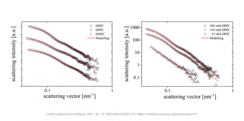
Apart from studies of the bilayer morphology, SAXS can be used for the size characterization of liposomes in the range from a few nanometers to a few hundred nanometers, depending on the type of liposome and instrument. In a recent publication from the Karlsruhe Institute of Technology (KIT) [10], SAXS was used to investigate the use of liposomes as a means of stabilizing fluorocarbons-based emulsions. In this foundational research, phospholipids with various head sizes, chain lengths and different concentrations of the phospholipids were investigated. Firstly, SAXS unambiguously confirms the formation of liposomes, whereas this was not clear for freshly prepared samples from other measurement techniques. Secondly, SAXS is insensitive to gas bubbles in the samples that are introduced during production. Therefore, SAXS can provide a more reliable measure for the size of the liposomes than the intensity weighted mean hydrodynamic size as measured by photon correlated spectroscopy (PCS). In this particular case, PCS measurements are further hindered by the small difference in the refractive indices of both phases, this is not a problem for SAXS as the contrast relies on differences in electron density rather than refractive index.
As can be seen in the images below, the radially averaged scattering patterns of the liposomes with different phospholipids (left) and at different concentrations (right) can be modeled with high quality fits. The results indicate that the size of the liposomes increases with increasing length of the phospholipid tail from 48 nm for DMPC to 59 nm for DSPC and 61 nm for DPPC. Varying the concentration of DPPC from 25 to 300 mM results in the formation of liposomes from 61 nm to well over the detection limit of the instrument. Furthermore, the increase in scattering intensity leads to the conclusion that the more phospholipids are present, the more liposomes can be formed.

These results present an important first step in the development of fluorocarbon-in-water emulsions which can be used for pharmaceutical purposes. Fluorocarbons have recently gained interest as carriers for hydrophobic drugs as well as gaseous oxygen. Furthermore, fluorocarbons find use as MRI contrast agents. Until now, the production process is far from optimal though, wasting a lot of the active ingredients – a problem which might be solved with the use of liposomes.
Xenocs SAXS solutions for R&D and manufacturing of drug carriers
Liposomes, among other nanoparticles, are attractive vehicles for drug payload delivery, providing endless possibilities for tackling various pharmaceutical needs. At Xenocs we provide solutions for the effective development of liposomes and other nanoparticles. Our products reduce the R&D phase by providing robust and reliable results. Moreover, they are well suited for manufacturing monitoring and quality control. This helps to ensure that safe products reach the market faster.
In Vietnam, please do not hesitate to contact us for more information and consultation for any of your applications with this beautiful product from #Xenocs.
References and Further Reading
- Sreij, R.; Dargel, C.; Moleiro, L. H.; Monroy, F.; Hellweg, T. Aescin Incorporation and Nanodomain Formation in DMPC Model Membranes. Langmuir 2017, 33 (43), 12351–12361. https://doi.org/10.1021/acs.langmuir.7b02933.
- Chaves, M. A.; Oseliero Filho, P. L.; Jange, C. G.; Sinigaglia-Coimbra, R.; Oliveira, C. L. P.; Pinho, S. C. Structural Characterization of Multilamellar Liposomes Coencapsulating Curcumin and Vitamin D3. Colloids and Surfaces A: Physicochemical and Engineering Aspects 2018, 549, 112–121. https://doi.org/10.1016/j.colsurfa.2018.04.018.
- Battista, S.; Marsicano, V.; Arcadi, A.; Galantini, L.; Aschi, M.; Allegritti, E.; Del Giudice, A.; Giansanti, L. UV Properties and Loading into Liposomes of Quinoline Derivatives. Colloids and Interfaces 2021, 5 (2), 28. https://doi.org/10.3390/colloids5020028.
- Borro, B. C.; Toussaint, M. S.; Bucciarelli, S.; Malmsten, M. Effects of Charge Contrast and Composition on Microgel Formation and Interactions with Bacteria-Mimicking Liposomes. Biochimica et Biophysica Acta (BBA) – General Subjects 2021, 1865 (4), 129485. https://doi.org/10.1016/j.bbagen.2019.129485.
- Lim, S. W. Z.; Wong, Y. S.; Czarny, B.; Venkatraman, S. Microfluidic-Directed Self-Assembly of Liposomes: Role of Interdigitation. Journal of Colloid and Interface Science 2020, 578, 47–57. https://doi.org/10.1016/j.jcis.2020.05.114.
- Wood, I.; Albano, J. M. R.; Filho, P. L. O.; Couto, V. M.; de Farias, M. A.; Portugal, R. V.; de Paula, E.; Oliveira, C. L. P.; Pickholz, M. A Sumatriptan Coarse-Grained Model to Explore Different Environments: Interplay with Experimental Techniques. Eur Biophys J 2018, 47 (5), 561–571. https://doi.org/10.1007/s00249-018-1278-2.
- BioXolver application note ANBX06 Probing the effect of oxidation on model lipid bilayers.
- Schilt, Y.; Berman, T.; Wei, X.; Barenholz, Y.; Raviv, U. Using Solution X-Ray Scattering to Determine the High-Resolution Structure and Morphology of PEGylated Liposomal Doxorubicin Nanodrugs. Biochim Biophys Acta 2016, 1860 (1 Pt A), 108–119. https://doi.org/10.1016/j.bbagen.2015.09.012.
- Nordström, R.; Zhu, L.; Härmark, J.; Levi-Kalisman, Y.; Koren, E.; Barenholz, Y.; Levinton, G.; Shamrakov, D. Quantitative Cryo-TEM Reveals New Structural Details of Doxil-Like PEGylated Liposomal Doxorubicin Formulation. Pharmaceutics 2021, 13 (1), 123. https://doi.org/10.3390/pharmaceutics13010123.
- Ullmann, K.; Meier, M.; Benner, C.; Leneweit, G.; Nirschl, H. Water-in-Fluorocarbon Nanoemulsions Stabilized by Phospholipids and Characterized for Pharmaceutical Applications. Advanced Materials Interfaces 2021, 8 (1), 2001376. https://doi.org/10.1002/admi.202001376
[i] https://dx.doi.org/10.3390%2Fpharmaceutics9020012
[ii] https://ec.europa.eu/health/documents/eudralex/vol-4_en
[iii]http://www.fda.gov/downloads/Drugs/GuidanceComplianceRegulatoryInformation/Guidances/ucm070570.pdf


